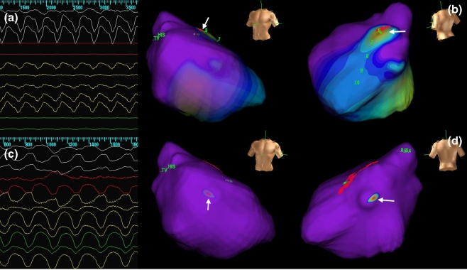
Fig. 2.
Electrocardiograms and endocardial activation maps in a patient having VTs with two different morphologies and cycle lengths. One VT was ablated successfully in the RVOT region, whereas the other one was seen to originate more apically in the RV. VT ventricular tachycardia; RV right ventricular

