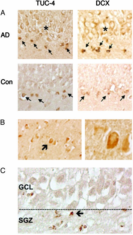
Fig. 3.
Immunohistochemical evidence for increased neurogenesis in hippocampus of AD brains. (A) TUC-4 and DCX are expressed in the SGZ of control and AD brain (arrows), but only in AD are large numbers of TUC-4- and DCX-immunopositive cells observed in GCL (*). Immunoreactive cells in the SGZ show shrunken cytoplasm and condensed nuclei, which is consistent with death of cells that do not transit to the GCL; a similar finding was observed in aged control brains (data not shown). (B) DCX-immunopositive cells (arrow) can also be detected in CA1 (Left, low power; Right, high power) of AD hippocampus. (C) Cells in SGZ express the 10-kDa caspase-8 cleavage product (arrow), suggesting caspase-dependent programmed cell death.

