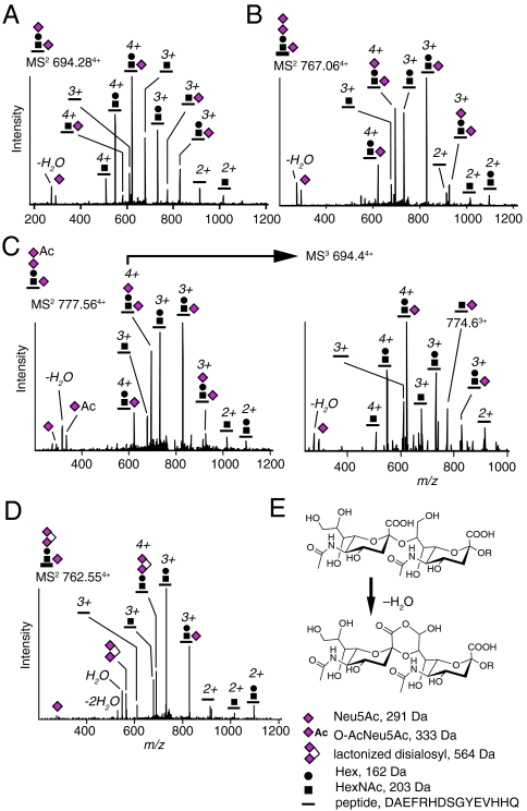
Fig. 1.
Glycosylated and sialylated Aβ1-15 peptides from human CSF. CID MS2 spectra of (A) SA2-Aβ1-15 and (B) SA3-Aβ1-15. (C) O-AcSA3-Aβ1-15 (Left) with MS3 fragmentation at m/z 694.4 (Right), and (D) lactonized SA3-Aβ1-15. (E) Structure of α2,8-linked disialic acid terminal and its lactonized form. The Neu5Ac oxonium ion and its loss of H2O are present at m/z 292 and 274, respectively. Neu5Ac, N-acetyl-5-neuraminic acid; Hex, hexose; HexNAc, N-acetylhexosamine; O-Ac, O-acetyl.

