Fig. 5.
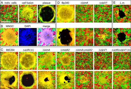
Intercellular spread and plaque formation occur through cell fusion. (A) Progression of events leading to plaque formation. (Left) HEK293-RFP (red) and -GFP (green) cells immediately after infection. Twelve hours later, a MNGC is formed (yellow, Center), which undergoes lysis and forms a plaque at 24 h (Right). (Scale bar, 500 μm.) (B) A MNGC (Left) was stained with DAPI (blue, Center). (Right) Triple-color merged image. (C) MNGC and plaque formation by BtE264 and mutants on HEK293 RFP + GFP monolayers 12 h (Upper) or 24 h (Lower) following infection or nanoblade delivery (“n”, ΔsctN, ΔsctNΔclpV1). (D) MNGC and plaque formation 12 h (Upper) and 24 h (Lower) after infection with Bp340 and mutants. (E) Plaque formation 56 h following infection with L. monocytogenes 10403S. The slight appearance of yellow at the edge of plaques is due to physical overlap of red and green cells and does not indicate cell fusion. Images are representative of multiple independent experiments.

