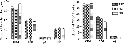
FIG. 1.
The relative percentages of total CD4+ T cells, CD8+ T cells, NK cells, and γδ T cells in the lamina propria of the small-intestinal mucosa were comparable in patients with T1D, HC subjects, and patients with CD. Lamina propria cells were isolated from the small-intestinal mucosa; simultaneously stained with Pacific Blue–conjugated anti-human CD4, APC-conjugated anti-human NKG2D, FITC-conjugated anti-human TCRδ1, and AmCyan-conjugated anti-human CD3 monoclonal antibodies; and FACS analyzed. Means of percentages ± SEM of CD4+ T cells (CD3+CD4+ cells), CD8+ T cells (CD3+CD8+ cells), γδ T cells (CD3+ TCRδ1+ cells), and NK cells (CD3− NKG2D+ cells) out of total lamina propria lymphocytes (left) and the means of percentages ± SEM of CD4+ T cells, CD8+ T cells, and γδ T cells out of CD3+ T cells (right) are indicated.

