FIG. 3.
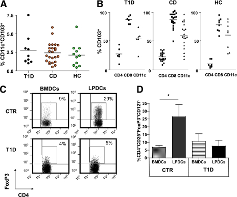
CD103+CD11c+ LPDCs are present in the small intestine of T1D patients but fail to induce FoxP3+ Treg cell differentiation. A: Percentages of CD103+CD11c+ LPDCs in the gut mucosa of patients with T1D, HC subjects, and patients with CD. Cells were gated on the DC region (see Supplementary Fig. 4). B: Percentages of cells that express the gut receptor CD103 among various gut lymphocyte subsets (CD4+ T cells, CD8+ T cells, and CD11c+ DCs) in the three groups. C: The capacity of small-intestinal CD11c+CD103+ LPDCs of T1D patients and control subjects (healthy or celiac individuals [CTRs]) to induce CD4+CD25+FoxP3+CD127− Treg cell differentiation was evaluated in conversion assays. BMDCs or small-intestinal CD11c+CD103+ LPDCs were obtained from T1D patients and CTRs and added at 1:10 ratio (DC to T cells) to autologous naive CD4+CD25− T cells isolated from PBMC and stimulated with soluble anti-human CD3 monoclonal antibody in the presence of rhIL-2 and rhTGF-β. Cells were collected after 4 days and FACS analyzed to measure percentages of CD4+CD25+FoxP3+CD127− Treg cells. One representative experiment out of three (T1D) or four (CTR) is shown (cells gated on CD4+CD25+CD127− cells). D: Means ± SEM of CD4+CD25+FoxP3+CD127− Treg cell percentages obtained in conversion assays with BMDCs or LPDCs in three (T1D) or four (CTR) independent experiments are shown. *P = 0.028. (A high-quality color representation of this figure is available in the online issue.)

