FIG. 1.
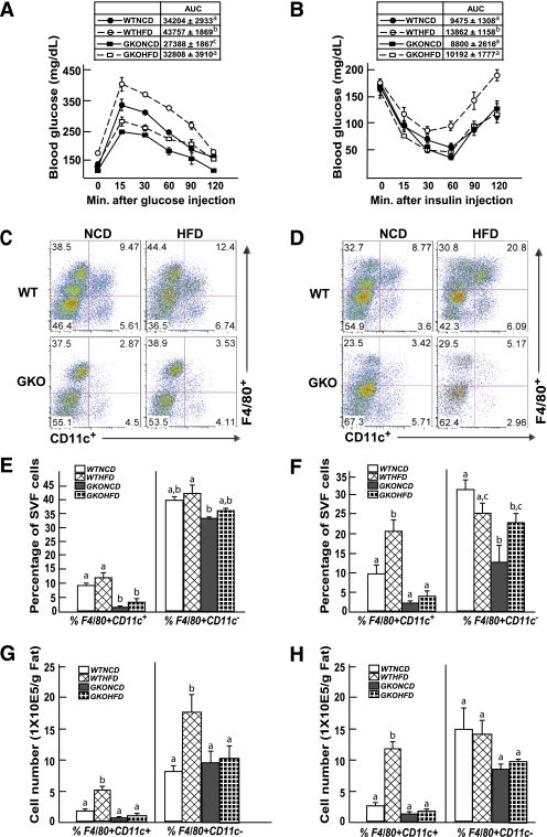
GM-CSF–null mice are protected against HFD-induced glucose and insulin intolerance and against HFD-induced adipose tissue macrophage infiltration. Wild-type and GM-CSF–null 9-week-old male mice were fed NCD or HFD for 4 weeks, were fasted for 16 h (IPGTT) or 4 h (ITT), and blood was drawn for determination of fasting glucose and insulin levels. The mice were given an intraperitoneal injection of 1 g/kg glucose (A) or 1 unit/kg insulin (B), and blood glucose and insulin levels were determined at 15, 30, 60, 90, and 120 min. WTHFD, wild-type mice fed the HFD; WTNCD, wild-type mice fed the NCD; GKOHFD, GM-CSF–null mice fed the HFD; GKONCD, GM-CSF–null mice fed the NCD. Area under the curve (AUC) for each condition (n = 4 mice per group) is shown in the insert. Identical letters indicate values that are not statistically different from each other (P > 0.05). Wild-type and GM-CSF–null 9-week-old male mice were fed the NCD or HFD for 4 (C, E, G) or 8 (D, F, H) weeks. The stromal vascular fraction from an entire epididymal adipose tissue fat pad was isolated and subjected to flow cytometry analysis after labeling with F4/80 and CD11c antibodies, as described in research design and methods (C, D). The data obtained from three to five independent experiments were calculated for the percentage of F4/80+CD11c+ and F4/80+CD11c− (E, F) and the total number of F4/80+CD11c+ and F4/80+CD11c− cells (G, H). Data were analyzed as described in statistical analysis and are shown as the mean ± standard error of the mean. Identical letters indicate values that are not statistically different from each other (P > 0.05). (A high-quality digital representation of this figure is available in the online issue.)

