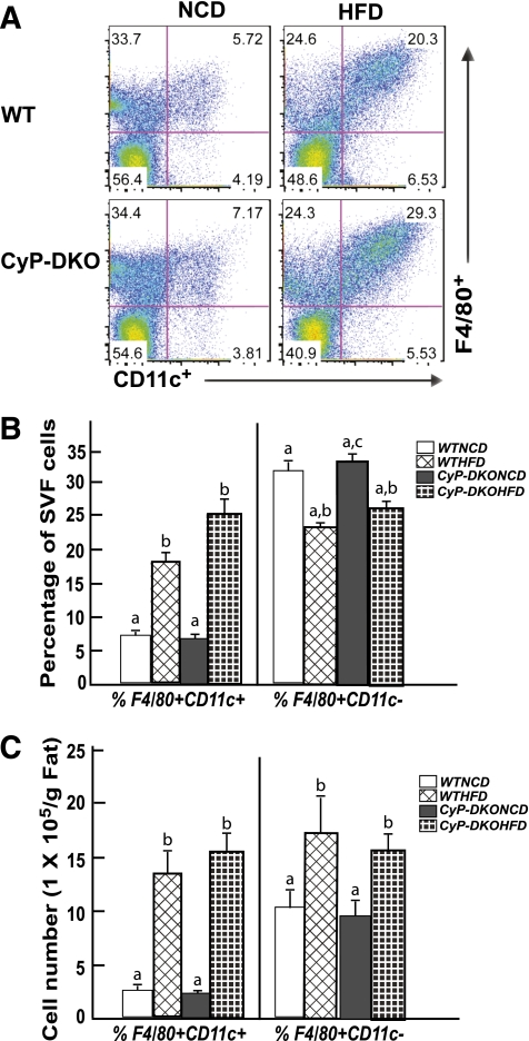
FIG. 7.
CyP-D–null (KO) mice display a normal extent of HFD-induced macrophage infiltration into adipose tissue. Wild-type and CyP-D–null 9-week-old male mice were fed the NCD or HFD for 12 weeks. The stromal vascular fraction (SVF) from an entire epididymal adipose tissue fat pad was isolated and subjected to flow cytometry analysis after labeling with F4/80 and CD11c antibodies as described in research design and methods. FACS analysis of SVF isolated from epididymal adipose tissue. B: Percentage of F4/80+CD11c+ and F4/80+CD11c− macrophages in SVF of epididymal adipose tissue. C: Total number of F4/80+CD11c+ and F480+CD11c− cells. Data shown are the mean ± standard error of the mean six mice per group and were analyzed as described in statistical analysis. Identical letters indicate values that are not statistically different from each other (P > 0.05).

