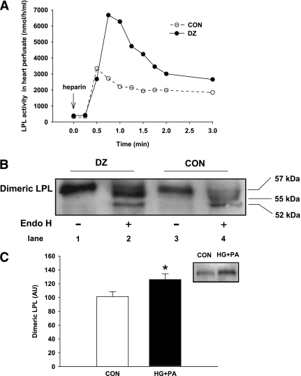
FIG. 2.
Increased LPL dimers are present in cardiomyocytes from DZ animals and cells acutely exposed to HG and PA. Four hours after injection of DZ, hearts were isolated and perfused with heparin (5 units/mL), and fractions of perfusate at the indicated times were analyzed for LPL activity (A). Subsequent to heparin perfusion and detachment of vascular LPL, heart homogenates were subjected to heparin–sepharose elution to isolate LPL dimers (in the 1.0 mol/L NaCl elutions). These fractions were combined, digested with (+) or without (−) endo H for 20 h, and concentrated by TCA precipitation, and LPL protein was determined by Western blot (B). Isolated cardiac myocytes were plated on laminin-coated 60 × 15-mm tissue culture dishes and treated with PA (1 mmol/L bound to 1% BSA) and 25 mmol/L glucose (HG+PA) for 2 h. Media containing 1% BSA was used as control (CON). Cellular dimeric LPL was determined by running cell lysates onto a heparin–sepharose column, and fractions eluted at 1.0 mol/L NaCl were used to determine LPL protein by Western blot (C). Results are the mean ± SE of five repeated experiments using different animals. *Significantly different from control, P < 0.05. AU, arbitrary units.

