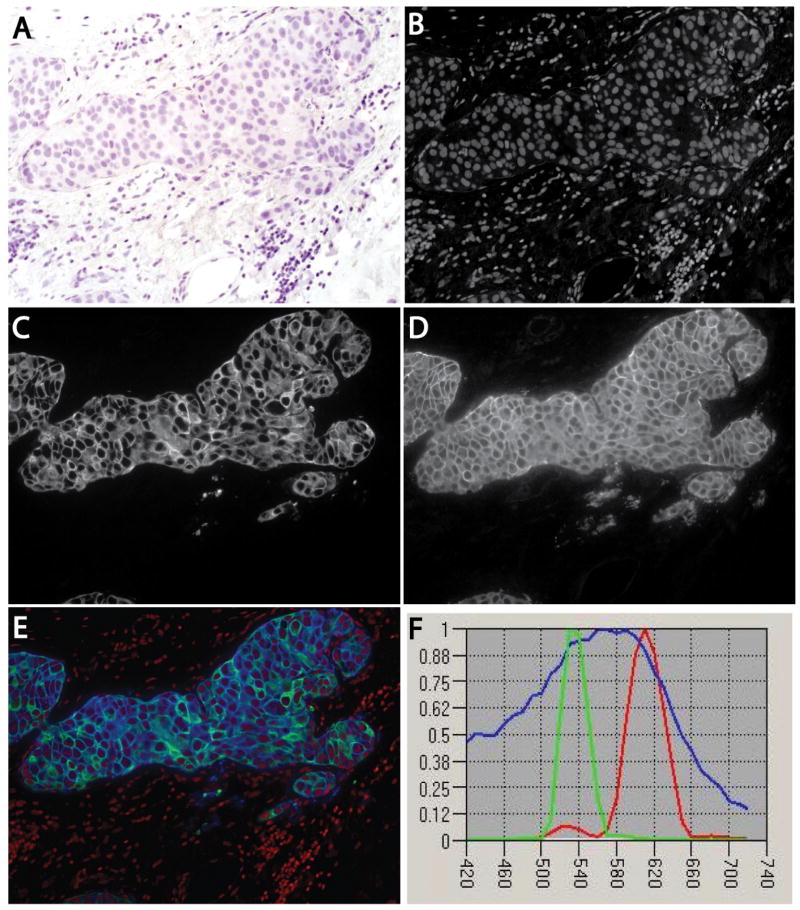
Figure 1.
Multiplex-stained human breast cancer specimen. A human breast cancer was stained for HER2 by immunofluorescence with Texas Red and for cytokeratin by immunofluorescence with Alexa-488, and counterstained with haematoxylin. The slide was imaged multispectrally in absorption and fluorescence modes, and the results were unmixed to yield non-overlapping channels. A, Brightfield image showing haematoxylin staining. B, Unmixed channel containing only cell nuclei, corresponding to the haematoxylin spectral signature. C, Unmixed channel for fluorescently stained cytokeratin. D, Unmixed channel corresponding to fluorescently stained HER2. E, Composite three-colour image with nuclei (red), cytokeratin (green), and HER2 (blue). F, Spectral signatures used for the unmixing computations, displayed using blue for haematoxylin (nuclei), green for Alexa-488 (cytokeratin), and red for Texas Red (HER2).

