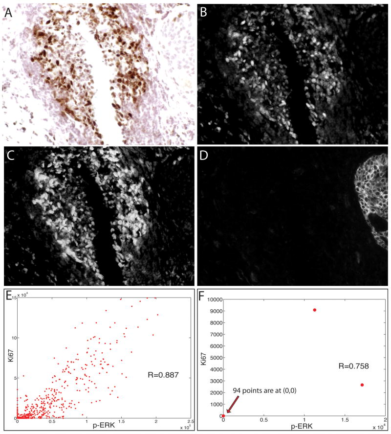
Figure 4.
Duplex analysis of phospho-extracellular signal-related kinase (p-ERK) and Ki67 immunostaining in lymphoid cells in a human breast carcinoma. A section of a breast tumour was stained sequentially with anti-p-ERK (SG Blue), anti-Ki67 [3,3-diaminobenzidine (DAB)] and anti-CK (Alexa-488) antibodies, and this was followed by haematoxylin staining, multispectral imaging (×400), and cytometric analysis. The brightfield image of a lymphoid nodule in the tumour is shown (A), along with the unmixed channels for DAB (Ki67) (B), SG Blue (p-ERK) (C), and Alexa-488 (cytokeratin) (D). Scatter plots of p-ERK (x-axis) and Ki67 (y-axis) staining intensity are shown for cells in the lymphoid nodule (E) and for tumour cells (F), with each dot representing one cell.

