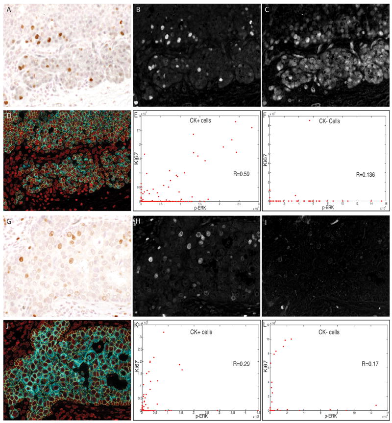
Figure 5.
Duplex analysis of phospho-extracellular signal-related kinase (p-ERK) and Ki67 immunostaining in human breast carcinoma cells. Sections of two different breast tumours were stained and analysed as described for Figure 5. Brightfield images of the two different tumours are shown (A,G), with the unmixed channels for 3,3-diaminobenzidine (Ki67) (B,H) and SG Blue (p-ERK) (C,I). Composite images showing whole-cell segmentation of the tumour (cytokeratin-positive) cells are shown (D,J). Scatter plots of p-ERK (x-axis) and Ki67 (y-axis) staining intensity are shown for tumour cells (E,K) and for non-tumour (stromal) cells (F,L), with each dot representing one cell.

