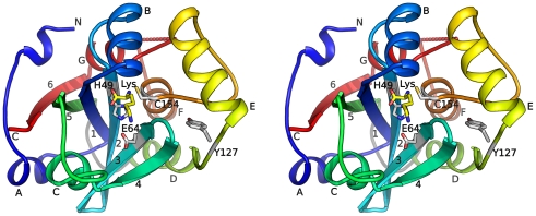
Figure 1. Crystal structure of BcPPNE.
Stereoview of ribbon representation of BcPPNE is color coded from blue (N-terminus) to red (C-terminus). The α-helices are labeled A to G, and β-strands 1 to 6. The bound lysine and active site catalytic residues are shown as sticks. The loop that unites the circular permuted sub-domains (residues 103–110) is colored grey. The 310 helices are not labeled.

