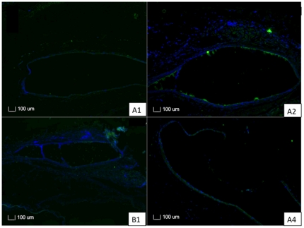
Figure 10. Sections of PLGA50/50 and PLC70/30 with immunohistochemistry stains for CD45 T cells.
Arrows point at the implant site. A, B: 1– Section of eye implanted with PLGA50/50 microfilm at 3 and 6 months respectively. A, B: 2 – Section of eye implanted with PLC70/30 microfilm at 3 and 6 months respectively.

