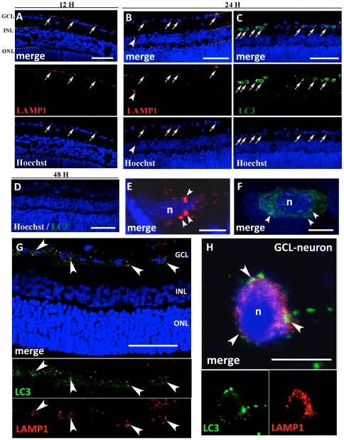
Figure 2. Immunofluorescence against LAMP1 and LC3.
All sections were counterstained with bisbenzimide (Hoechst staining) to show retinal layers. Twelve hours after I/R, LAMP1 positive cytoplasmic granules are present (A) and 24 h after I/R both LAMP1 (B) and LC3 (C) positive granules are present in the GCL (arrow) and INL (arrowheads). LC3-positive vesicles are more represented than LAMP1 vesicles. LC3-immunopositivity is absent after 48 h (D). At high magnification (E–F), clear cytoplasmic lysosomal LAMP1 positive vesicles (arrowheads) can be observed in the GCL at 24 h from the insult (E). LC3 labelling appear as numerous fluorescent dots (arrowheads) after 24 h from the I/R (F). Double immunolabeling against LAMP1 (red) and LC3 (green) at 24 h shows the relationship between autophagosomes and lysosomes (G). The increase in punctuate LC3 and LAMP1 occurs in the same neurons after 24 h (G, arrowheads). At high magnification, confocal microscopy reveals that autophagic marker and LAMP1 colocalize, suggesting that fusion of autophagosomes with lysosomes occur in dying neurons after I/R (arrowheads, H). Abbreviations as in Figure 1. Scale bars = 50 µm (A); 100 µm (B, C, D and G) and 5 µm (E, F and H).

