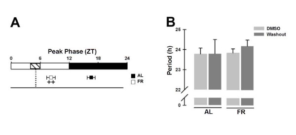
Figure 2.
Per1-luc bioluminescence rhythms in liver explants treated with DMSO. (A) Peak phases (in ZT) from Per1-luc expression rhythms calculated from the first peak occurring at the beginning of the culture. The dashed rectangle inside the subjective light period indicates the former food-restricted schedule, and the vertical dotted line represents the former meal time of the FR rats. (B) Period from Per1-luc expression in liver cultures from AL (left bars) and FR (right bars) rats during and after (washout) DMSO treatment. All data plotted are presented as means ± SEM, n = 6. ++Significant difference between AL vs. FR group on the indicated treatment day (Bonferroni post hoc test, α = 0.05).

