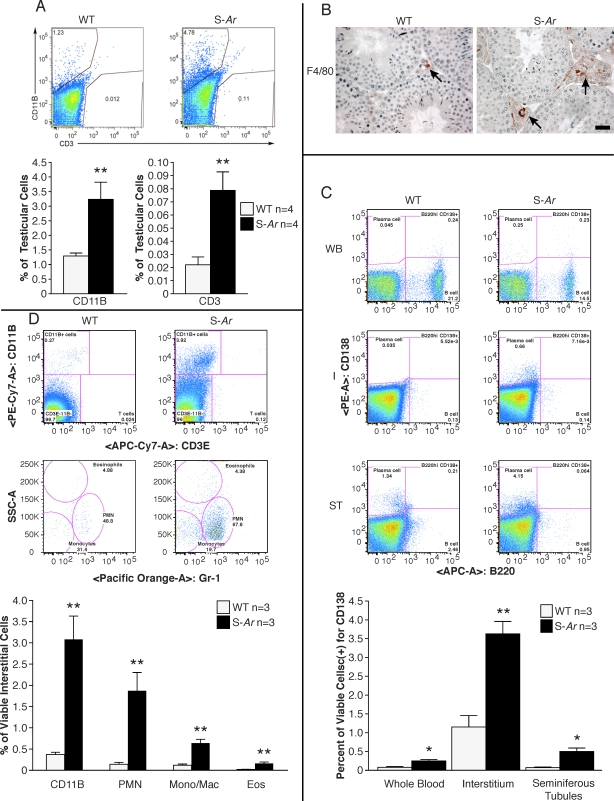
FIG. 4.
Inflammatory cells infiltrate the testes of S-Ar mutant mice. A) Flow cytometric analysis of whole testicular cell suspensions from WT and mutant mice show significantly higher percentages of CD11B-positive macrophages, neutrophils, and eosinophils and CD3-positive T lymphocytes in S-Ar mutant mice (both n = 4). An unstained control was used to exclude negative testicular cells and set gates for the CD11B and CD3 populations. B) Immunohistochemistry of macrophages stained with F4/80 antibody. Positive cells are stained red in the interstitium of WT and S-Ar mutant testes and noted with arrows. Bar = 20 μm (applies to both images). C) In comparison to WT, percentages of CD138(+) plasma cells in whole blood, testicular intersitium, and seminiferous tubules were significantly higher in S-Ar mutant mice (all n = 3). D) In comparison to WT, the interstitia of S-Ar mutants contained significantly higher percentages of CD11B(+) cells. It was possible to subdivide the CD11B-positive population into neutrophils, monocytes, and eosinophils based on costaining with anti-GR-1 antibody and by side scatter to detect granularity (all n = 3). *P < 0.05; **P < 0.01.

