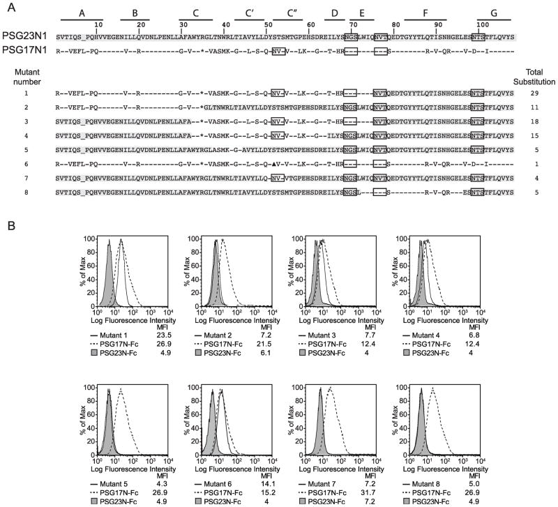
Figure 5.
PSG-mutants and mapping of the CD9-binding site in the N1-domain of PSG17. (A) Identity between amino acids in the N1 domains of PSG17 and PSG23 is shown with a dash. Potential glycosylation sites are boxed. Highlighted amino acids indicate correspondence to the PSG23N1 sequence. “*” represents the K35 to R substitution in PSG17 required to introduce a restriction site and “▲” indicates the N52 to A mutation in mutant 6, which deletes one of the potential N-linked glycosylation sites in PSG17. The β-strands (A to G) are indicated with lines on the upper part of the figure. Total substitution refers to the number of amino acids changed from the wild type sequence. (B) Sog9 expressing murine CD9 were treated with 30 μg/ml of the mutants shown in part A, PSG17N-Fc or PSG23N-Fc followed by PE-conjugated anti-human Fcγ.

