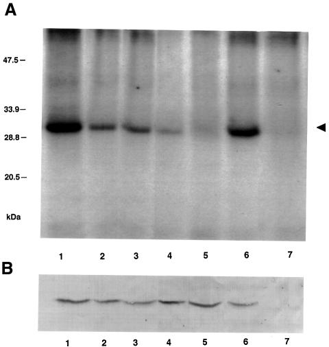
Fig. 1. MP phosphorylation in vivo. (A) PhosphorImage of MP immunopurified from plant cell walls and analyzed by SDS–PAGE as described in Materials and methods. Transgenic plants expressed the following MP derivatives: lane 1, wild-type MP; lane 2, sb-10; lane 3, sb-11; lane 4, sb-12; lane 5, sb-13. MP phosphorylation during TMV infection: lane 6, cell walls of TMV-infected plants; lane 7, cell walls of uninfected plants. The numbers on the left indicate molecular mass standards in thousands of daltons. An arrowhead indicates the position of TMV MP. (B) Western blot analysis of MP amounts used in the experiment described in (A).

An official website of the United States government
Here's how you know
Official websites use .gov
A
.gov website belongs to an official
government organization in the United States.
Secure .gov websites use HTTPS
A lock (
) or https:// means you've safely
connected to the .gov website. Share sensitive
information only on official, secure websites.
