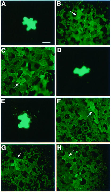
Fig. 3. Host-dependent effect of mimicking MP phosphorylation on plasmodesmal permeability. (A and E) Fluorescently labeled dextran (10 kDa) microinjected alone into N.tabacum or N.benthamiana, respectively. (B and F) Wild-type MP mixed with 10 kDa fluorescently labeled dextran and microinjected into N.tabacum or N.benthamiana, respectively. (C and G) del 7 mixed with 10 kDa fluorescently labeled dextran and microinjected into N.tabacum or N.benthamiana, respectively. (D and H) sb-9 mixed with 10 kDa fluorescently labeled dextran and microinjected into N.tabacum or N.benthamiana, respectively. Intercellular distribution of the signal was visualized 25–45 min after injection. Arrows indicate injected cells. Magnification: 120×. Note that black spaces between cells represent intercellular air pockets characteristic for tobacco leaf mesophyll tissues (Waigmann et al., 1994).

An official website of the United States government
Here's how you know
Official websites use .gov
A
.gov website belongs to an official
government organization in the United States.
Secure .gov websites use HTTPS
A lock (
) or https:// means you've safely
connected to the .gov website. Share sensitive
information only on official, secure websites.
