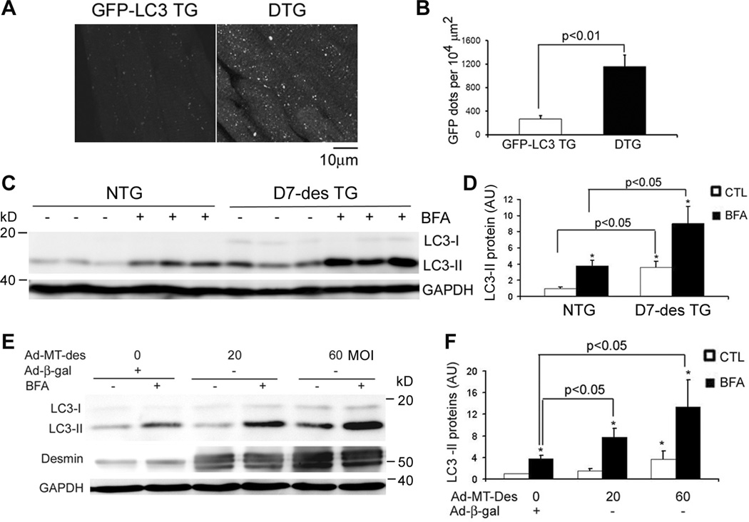
Figure 1.
Expression of a DRC-linked mutant desmin increases autophagic flux in mouse hearts and NRVMs. A and B, Confocal microscopic analysis of GFP-LC3 distribution in ventricular myocardium. D7-des tg mice were cross-bred with GFP-LC3 mice. The resultant GFP-LC3::D7-des double tg (DTG) mice and their littermate GFP-LC3 single tg mice were analyzed at 2 months of age. The representative images (A) and the quantitative analysis of the number of GFP-LC3 puncta (B) are presented. The GFP dot data (B) were quantified from the 3 randomly selected fields per section, 3 representative sections per heart, and 3 hearts per group. C and D, Increases in autophagic flux in the D7-des tg mouse heart. D7-des tg and NTG littermate mice at 2 months were intravenously injected with one dose of bafilomycin A1 (BFA, 6µmol/kg) or vehicle control (CTL) at 3 hours before the hearts were harvested for western blot analyses of LC3. Representative images (C) and a summary of LC3-II densitometry data (D) are presented. *p<0.05 vs. CTL. E and F, Autophagic flux was increased in NRVMs expressing DRC-linked mutant desmin (MT-Des). Cultured NRVMs were infected with Ad-MT-Des or Ad-β-gal (as control). Six days after infection, the cells were treated with BFA (100nM) or DMSO for 3 hours before being harvested for western blot analysis for the indicated proteins. Representative images (E) and a summary of densitometry data (F) are presented. *p<0.05 vs. CTL; #p<0.05 vs. Ad-β-gal/CTL; N=3/group in all cases. AU = arbitrary units.

