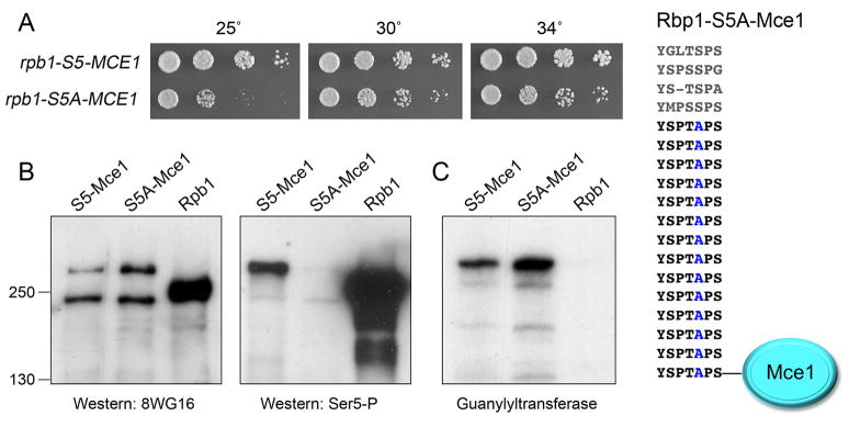
Figure 3. The lethality of S5A is bypassed by fusion of capping enzyme to the CTD.
(A) The S5A-Mce1 fusion is depicted at right. rpb1-S5-MCE1 and rpb1-S5A-MCE1 cells were grown in liquid culture at 30°C and serial dilutions were spotted to YES agar. The plates were photographed after 4 d at 25°C, or 3 d at 30 and 34°C. (B) Pol II immunoblots of extracts of rpb1+, S5-MCE1, and S5A-MCE1 cells using 8WG16 (left panel) or anti-Ser5-P (right panel) antibodies. The positions and sizes (kDa) of marker proteins are indicated at left. (C) Guanylyltransferase activity was gauged by label transfer from [α32P]GTP to the active capping enzymes in the extract to form covalent enzyme-[32P]GMP adducts detectable by SDS-PAGE and autoradiography. The positions of marker proteins are as in panel B. (See also Fig. S1.)

