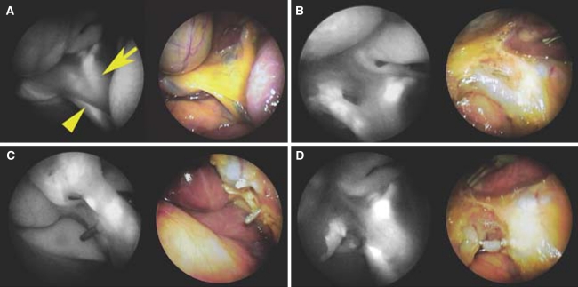
Fig. 3.
Near infrared fluorescen cholangiography during laparoscopic cholecystectomy [78]. A Cystic duct running parallel to common hepatic duct, B isolation of cystic duct from anterior side of Calot’s triangle, C isolation of cystic duct from posterior side of Calot’s triangle, D closure of cystic duct (with kind permission from John Wiley and Sons Ltd © 2010, all rights reserved)

