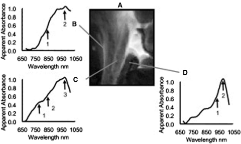
Fig. 4.
Hyperspectral cholangiography. A Near-infrared (NIR) laparoscopic hyperspectral image of the hepatoduodenal ligament in live anesthetized pigs. B An artery indicated by spectra with broad oxyhemoglobin peak and a small water peak at 970 nm. C A vein is identified by spectra containing a deoxyhemoglobin shoulder, a broad oxyhemoglobin peak, and a small water peak. D The common bile duct is associated with spectra containing a lipid shoulder and a prominent water peak [82] (with permission from Elsevier Inc., © 2008)

