
Figure 2.
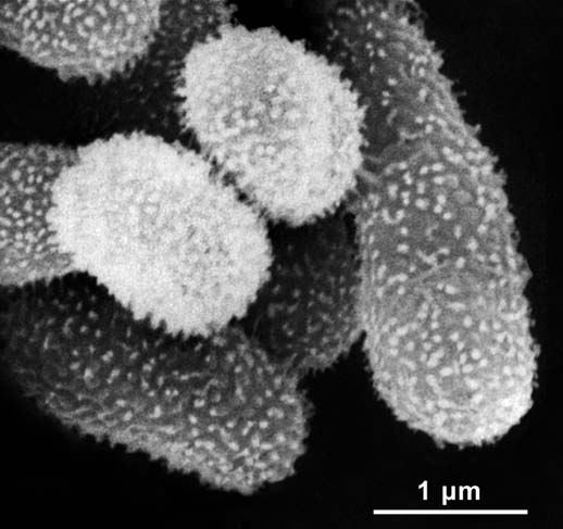
Scanning electron micrograph of a cluster of cells of Methylobacterium funariae F3.2T. The microbes were isolated from the upper side of a moss phylloid as shown in Figure 1. The bacteria depicted here grew on a protonema cell of the moss F. hygrometrica before the sample was fixed and prepared for microscopic examination.