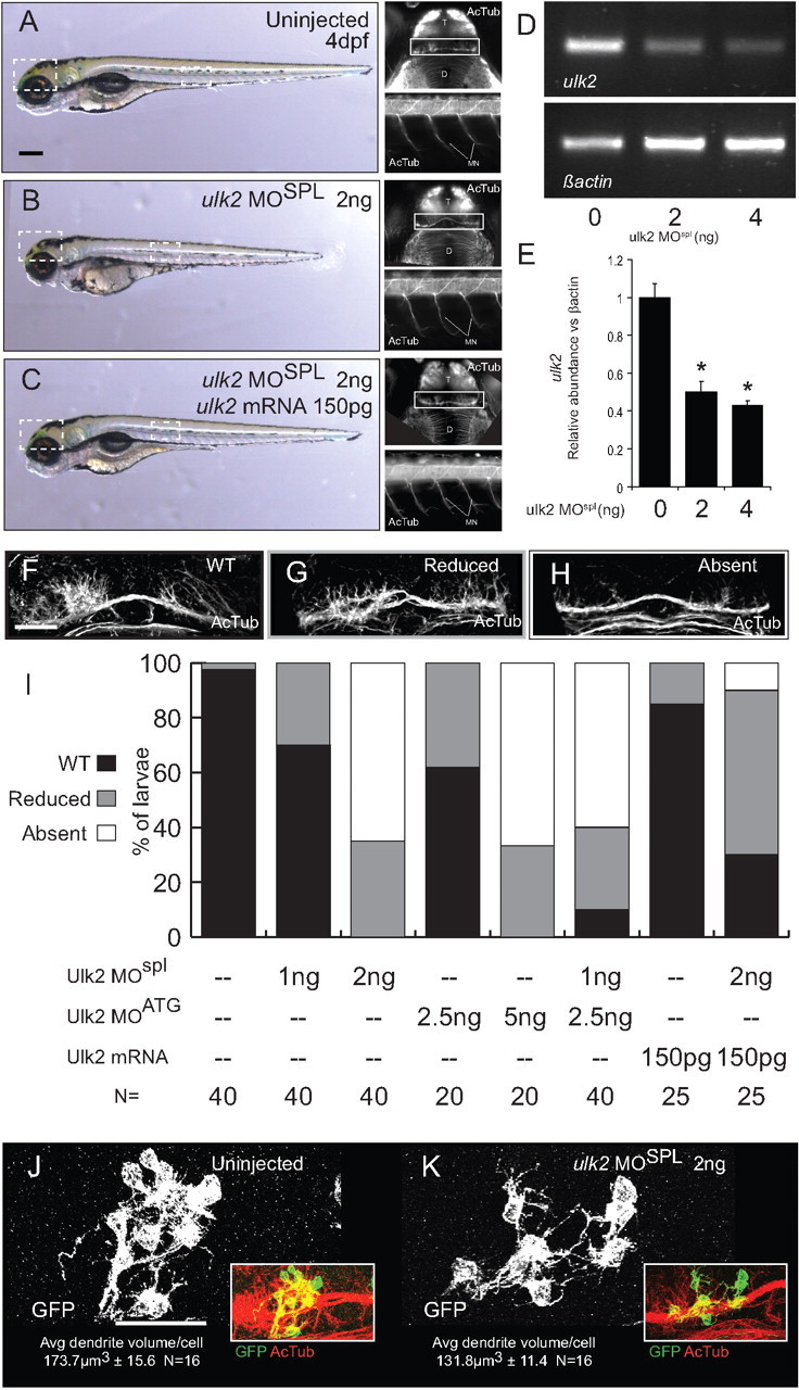
Figure 4.

Antisense knockdown of Ulk2 inhibits the formation of Hb neuropil. A, B, Injection of 2 ng of ulk2MOspl at the one-cell stage produces a mild decrease in head size and body length at 4 dpf. The general organization of the central (top insets) and peripheral (bottom insets) nervous system as revealed by acetylated tubulin immunofluorescence is intact in morphant larvae (D, diencephalon; T, telencephalon; MN, motor neurons), but Hb neuropil development (white boxes) is disrupted. C, Injection of prespliced Ulk2 mRNA at the one-cell stage rescues all the phenotypes of Ulk2 morpholino treatment. D, The ulk2 transcript is depleted by morpholino injection. RT-PCR with ulk2 primers (top panel) shows reduction of ulk2 mRNA relative to β-actin (bottom panel). E, Quantification of band intensity in RT-PCR replicates (0 ng = 1 ± 0.07 A.U., 2 ng = 0.5 ± 0.05 A.U., 4 ng = 0.43 ± 0.02 A.U., N = 3). F–H, Morphant larvae can be assigned to one of three categories based on the degree of Hb neuropil elaboration. I, When injected with either 2 ng of ulk2MOspl or 5 ng of ulk2MOATG, the formation of neuropil in the Hb is inhibited in most larvae (white bars), a phenotype not observed in injections of half-maximal dosage (1 and 2.5 ng, respectively). Hb neuropil inhibition was also observed when half-maximal doses of each MO were combined, indicating that both morpholinos target the same transcript. Injection of 150 pg of in vitro-transcribed ulk2 mRNA was able to rescue Hb neurite formation when coinjected with 2 ng of ulk2MOspl. J, K, Mosaic scatter labeling of Hb neurons with memGFP, followed by average dendrite volume quantification highlights changes in average dendrite volume between uninjected (J) and ulk2MO-treated (K) larvae. Scale bars: A, 100 μm; F, J, 50 μm.
