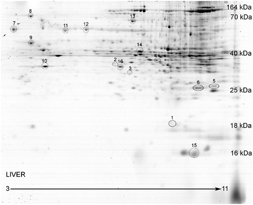
Figure 2.

Representative image of mouse liver proteins separated by 2D SDS-PAGE. The protein spot density of each spot was normalized to the total spot density of all spots on the gel. The proteins on the gel span an isoelectric focusing point of 3 to 11. The acidic proteins are on the left, while the basic proteins are on the right. The molecular weights of the liver protein spots are within the range of approximately 10 kDa and 170 kDa. Several proteins spots were identified to reference the molecular weights listed along the gel. There are varying intensities in the proteins spots on the gel image. The darker spots (or spots that are higher in intensity) are more abundant protein spots, while the lower intensity spots are lower abundance proteins.
