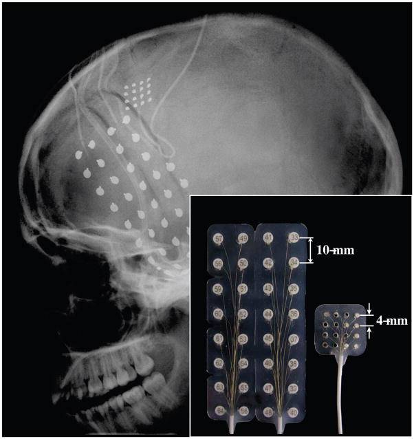
Fig. 1.
Head x-ray (lateral view) showing the locations and sizes of the implanted ECoG electrodes. One micro-ECoG grid (16 contacts), one regular ECoG grid (32 contacts), and two 6-contact regular ECoG strips were implanted. Inset: A side-by-side comparison of the regular ECoG grid and the micro-ECoG grid showing the center-to-center electrode spacing.

