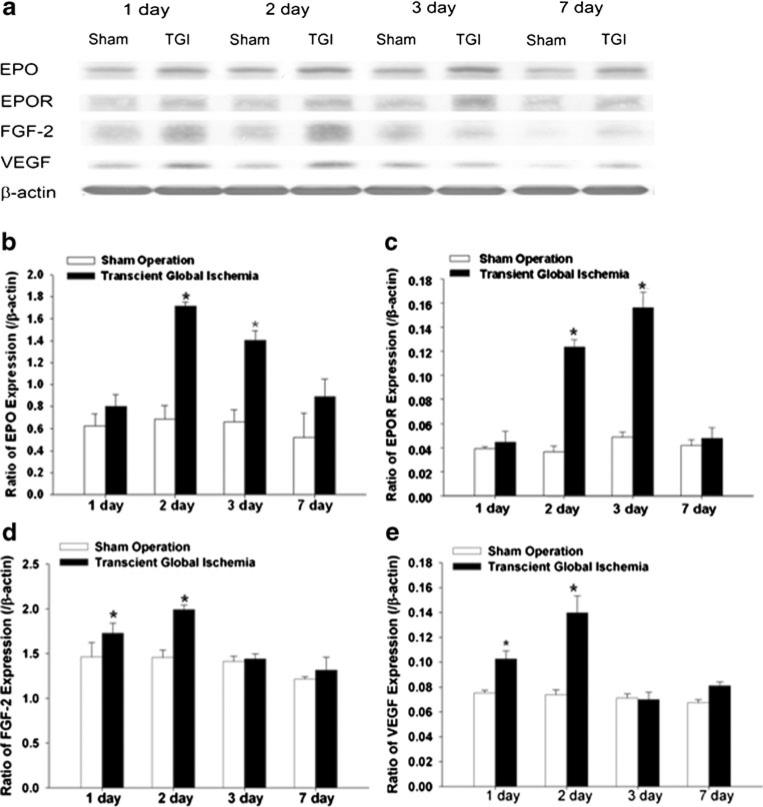
Fig. 3.
Upregulation of neurotrophic and neurogenetic factors after sublethal TGI. Western blot analysis was performed for both hemispheres to detect expression of EPO, EPOR, FGF-2, and VEGF protein levels at different days after TGI. a Representative Western blotting in the neocortex 1, 2, 3, and 7 days after sublethal TGI. Beta-actin was used as loading control. b–e Quantification of the expression of neurotrophic and neurogenetic factors EPO (b), EPOR (c), FGF-2 (d), and VEGF (e) at different days after the TGI. The gray intensity of corresponding bands was normalized to β-actin. N=3 independent assays in each group at each time point. *P<0.05 compared with controls

