Fig. 6.
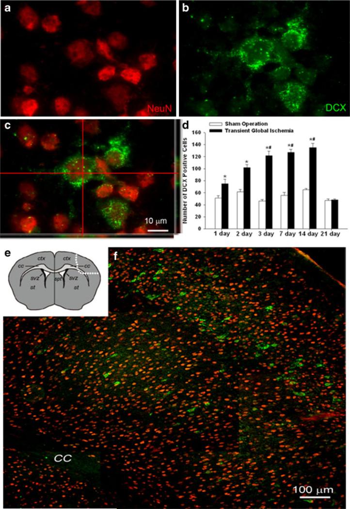
Sublethal global ischemia enhanced DCX-positive cells in the neocortex. Confocal imaging of NeuN and DCX double staining in the neocortex of the control and TGI brain. a, b NeuN (red) and DCX (green) labeling, respectively. c The confocal image shows overlapping of NeuN and DCX staining. Note the colocalization of red and green colors in the two side images. d Quantification of DCX-positive cells in both left and right hemispheres, counted 1–21 days after TGI and compared with sham groups. TGI significantly stimulated DCX expression. By 21 days after TGI, DCX expression subsided. *P<0.05 compared to control. N=6 in each group. e The diagram shows the area outlined by dotted line where the image f was taken from. f DCX-positive cells distributed in the neocortex in the layers I, III, and V. Bar=10 μm in a–c. Bar=100 μm in f

