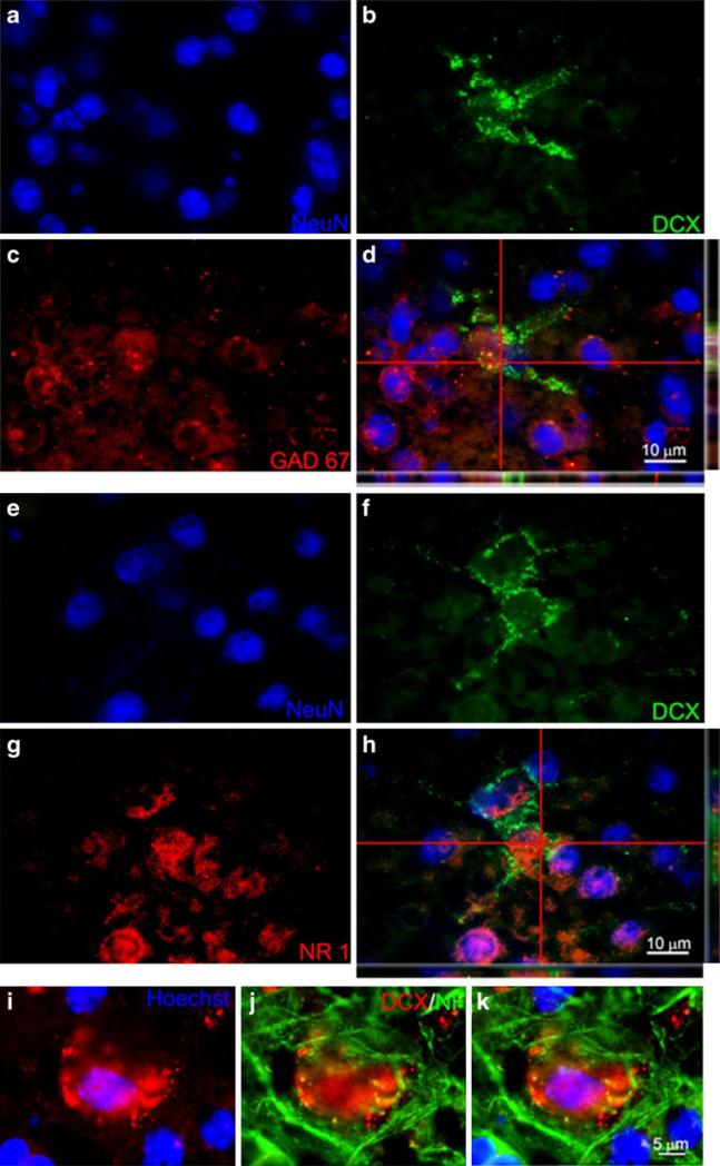
Fig. 7.
DCX-positive cells colocalized with mature neuronal markers. Confocal imaging analysis of colabeling of DCX and neuronal markers in the neocortex 7 days after TGI. a–c NeuN (blue), DCX (green), and GAD67 (red) staining, respectively. d The three-dimensional confocal image shows colabeling of NeuN and the GABAergic neuronal marker GAD67. Note the colocalization of green and red colors in side images. e–g NeuN (blue), DCX (green), and NR1 (red) staining, respectively. h The confocal image shows colocalization of DCX with the glutamatergic NMDA receptor gene NR1. i–k Enlarged images showed that DCX (red) colocalized with another mature neuronal marker neurofilament (NF; green). Bar=10 μm in a–h. Bar=5 μm in i–k

