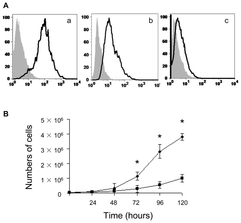
Figure 1. Immunophenotyping and high proliferation capacity of EPCs.
(A) Immunophenotyping of cell-surface by flow cytometry (a, CD-31; b, VE-cadherin; c, AC133). Shown are representative data from 4 independent experiments. Isotype controls are indicated in gray shadow. (B) Growth of EPCs (diamonds) and HAECs (squares) in EGM-2 was evaluated in equivalently seeded in vitro. *p<0.01.

