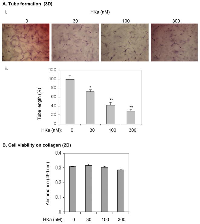
Figure 5. HKa inhibits EPC differentiation without affecting cell viability.
(A) i. Morphogenesis of EPCs in collagen gel. EPCs were suspended in EBM-2 containing 25 μM ZnCl2 and incubated with or without HKa on ice for 20 minutes. After mixture with the collagen gel solution, EPCs were cultured in the presence of VEGF (25 ng/mL) for 48 hours. ii. Tube length was measured and analyzed as described in the Figure 4A legend. Experiments were done in triplicate. * p<0.01; **p<0.005. (B) EPCs were cultured on collagen-coated plate in the presence of VEGF (25 ng/ml) plus HKa at the indicated concentrations for 48 hours (n=4). The cell number was determined as described in the Methods.

