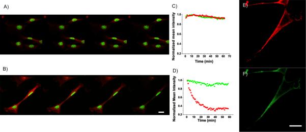
Fig. 2.
Confocal fluorescence imaging and photo-stability of C20 Ag nanodots (green) in NIH 3T3 cells co-labeled with HCS red cell stain (red) under one photon excitation (OPE, 458 nm, A) or two photon excitation (TPE, 720 nm, B). The photostability can be easily seen by the color decay of images in nuclei (green C20 Ag nanodots) and cytoplasm (HCS red cell stain), and illustrated in C (for OPE) and D (for TPE) by their mean fluorescence intensity of individual emitters in the corresponding scanning images. The green Ag nanodots show similar photostability to HCS red cell stain under OPE, but much greater stability under TPE. E and F, red and green channels from fixed NIH 3T3 cells co-stained with anti-OxoPhos/ATATC8 Ag nanodots (red) and anti-Heparin Sulfate/AATTC12 Ag nanodots (green). Scale bar, 30 μm.

