
Fig. 3.
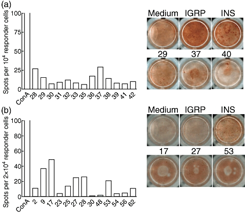
Low and variable responses against apoptosis-specific epitopes at 10 weeks of age. (a) Peptides 28–42 were screened, all of which are derived from myosin type A, except 28 (lamin B1). Background using medium only was seven spots. (b) Peptides 1–90 were screened, derived from actin cyto beta (1–8), hnRNP K (9–16), lamin B1 (17–28), myosin type A (29–88) and Rho GDI 2 (89–90). Only peptides associated with above-background detection are plotted. Concanavalin A stimulation was used as an additional positive control. Background using medium only was 57 spots in this experiment. Each experiment represents pooled expanded islet CD8 T cell populations from two mice. The entire peptide library was screened twice in four individual experiments using mice of the same age.