
Fig. 7.
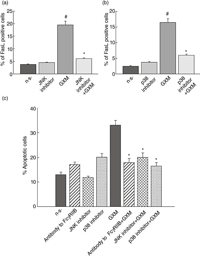
Evaluation of Fas ligand (FasL) surface up-regulation and glucuronoxylomamman (GXM)-induced apoptosis. (a,b) MonoMac6 cells (1 × 10⁶/ml), treated as described in Fig. 2, were stained with phycoerythrin (PE)-labelled monoclonal antibody (mAb) to FasL and percentage of FasL-positive cells was analysed by flow cytometry. Bars represent the mean ± standard error of the mean (s.e.m.) of three experiments. #P < 0·05 (GXM-treated versus untreated); *P < 0·05 [c-Jun NH2-terminal kinase (JNK) or p38 inhibitor GXM- treated, versus GXM-treated]. (c) MonoMac6 cells (1 × 10⁶/ml), treated as described in Fig. 5, were incubated with peripheral blood lymphocytes (PBL), pretreated with phytohaemagglutinin (PHA) as described in Materials and methods, at an effector : target ratio (E : T) = 10/1. The percentage of PBL undergoing apoptosis was quantified after 24 h of incubation by propidium iodide (PI) staining and analysed using a fluorescence activated cell sorter (FACScan) flow cytofluorometer. The PBL apoptosis rate resulted similar whether in the presence or absence of MonoMac not treated with GXM. Bars represent the mean ± s.e.m. of seven experiments. The incubation with irrelevant goat polyclonal immunoglobulin (Ig)G did not affect PBL apoptosis *P < 0·05 (antibody to FcγRIIB, or JNK or p38 inhibitors plus GXM-treated versus GXM-treated).