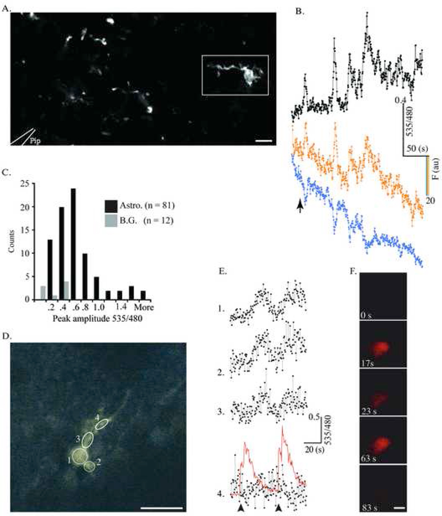
Figure 5.
YC 3.60-expressing glia respond to glutamate in situ. A. YC 3.60-containing astrocytes in a cortical slice (S100β-YC-C mouse) were imaged in a Zeiss Upright two-Photon microscope using non-descanned detectors. A picospritzer ejected 100nL of 500µM glutamate from a 1 MΩ resistance pipette tip (represented by white outlines at bottom left. Scale bar = 50 µm). In this focal plane, only one cell is visible and the rest of the fluorescent profiles are parts of cells. Images (512×256 pixels) were acquired at 2Hz (1.6µs pixel dwell time). B, The change in 535/480 during glutamate ejection (arrow) for the cell within the rectangle in A (black trace, top). The traces in color (cyan and yellow) are single wavelength traces at 480 and 535 nm respectively. C, Frequency histogram of compiled data from all the experiments (81 cells, 9 slices) shows YFP/CFP ratio change from baseline levels to peak amplitude upon glutamate addition (R/R0). B.G. denotes background. D– F, Cerebellar Bergmann glia were imaged and treated identically to the cortical astrocytes in A. Images (256×256 Pixels) were acquired at 2Hz (1.92 µs pixel dwell time). Circles delineate ROIs drawn over the cell where measurements were made (Scale bar = 20 µm). In this experiment, glutamate within the microinjection pipette was spiked with Quantum Dot 655 nanocrystals to allow direct visualization of the glutamate injection during imaging. E, YC 3.60 reports Ca2+ activity in ROIs 1–4 in D during two sequential glutamate injections. The arrow represents the 1st microinjection of 300µM glutamate, and the red trace represents the QD 655 fluorescence following injection in the ROI 4 trace. F. Time-lapse stills of the QD 655/Glutamate microinjection with time points labeled. The diameter of the QD cloud is roughly twice the diameter of the Bergmann glial cell body (Scale bar = 20 µm).

