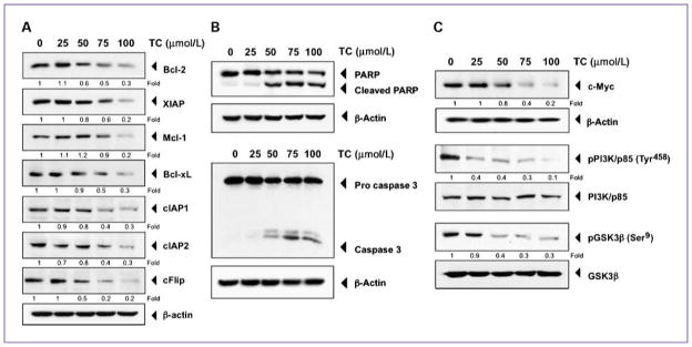
Fig. 2.
Thiocolchicoside induces apoptosis and inhibits proteins involved in cell proliferation. A, thiocolchicoside downregulates the expression of antiapoptotic proteins. KBM5 cells were treated with 25, 50, 75, and 100 μmol/L of thiocolchicoside for 24 h; whole-cell extracts were prepared and analyzed by Western blot using antibodies against Bcl-2, XIAP, Mcl-1, Bcl-xL, cIAP1, cIAP2, and cFlip. B, thiocolchicoside induces apoptosis. KBM5 cells were treated with 25, 50, 75, and 100 μmol/L of thiocolchicoside for 24 h. Whole-cell extracts were prepared and analyzed by Western blot using antibodies against PARP and caspase-3. C, thiocolchicoside downregulates the expression of proteins involved in cell proliferation. KBM5 cells were treated with 25, 50, 75, and 100 μmol/L of thiocolchicoside for 24 h. Whole-cell extracts were prepared and analyzed by Western blot using antibodies against c-Myc, the phosphorylated PI3K/p85, and phosphorylated GSK3β proteins. Unphosphorylated proteins of PI3K/p85 and GSK3β as well as β-actin were used as a loading control. Numbers below each panel indicate fold differences after normalization to β-actin.

