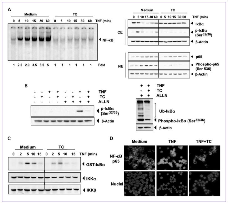
Fig. 5.
Thiocolchicoside inhibits TNF-dependent IκBα phosphorylation, IκBα degradation, p65 phosphorylation, and p65 nuclear translocation. A, thiocolchicoside inhibits TNF-induced activation of NF-κB. KBM-5 cells were incubated with 100 μmol/L thiocolchicoside for 24 h, treated with 0.1 nmol/L TNF for the indicated times, and then analyzed for NF-κB activation by EMSA (left). A, right, effect of thiocolchicoside on TNF-induced IκBα degradation, p65 phosphorylation, and p65 nuclear translocation. Cells were incubated with 100 μmol/L thiocolchicoside for 24 h and treated with 0.1 nmol/L TNF for the indicated times. Cytoplasmic extracts (CE) and nuclear extracts (NE) were prepared, fractionated on SDS-PAGE, and electrotransferred to nitrocellulose membrane. Western blot analysis was done using the indicated antibody. An anti-β-actin antibody was the loading control. B, effect of thiocolchicoside on the phosphorylation of IκBα by TNF. Cells were preincubated with 100 μmol/L thiocolchicoside for 24 h, incubated with 50 μg/mL ALLN for 30 min, and then treated with 0.1 nmol/L TNF for 10 min. Cytoplasmic extracts were fractionated and then subjected to Western blot analysis using a phospho-specific anti-IκBα antibody. An anti-β-actin antibody was used as loading control (left). B, right, thiocolchicoside inhibits ubiquitination of IκBα. Cells were preincubated with 100 μmol/L thiocolchicoside for 24 h, incubated with 50 μg/mL ALLN for 30 min, and then treated with 0.1 nmol/L TNF for 10 min. Cytoplasmic extracts were fractionated and then subjected to Western blot analysis using a phospho-specific anti-IκBα antibody. An anti-β-actin antibody used as the loading control. C, direct effect of thiocolchicoside on IKK activation induced by TNF. Whole-cell extracts were immunoprecipitated with antibody against IKKβ and analyzed by an immune complex kinase assay. To examine the effect of thiocolchicoside on the level of expression of IKK proteins, whole-cell extracts were fractionated on SDS-PAGE and examined by Western blot analysis using anti-IKKα and anti-IKKβ antibodies. D, immunocytochemical analysis of p65 localization. Cells were incubated with 100 μmol/L thiocolchicoside for 24 h and then treated with 1 nm TNF for 15 min. Cells were subjected to immunocytochemical analysis.

