Figure 1.
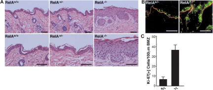
Hyperplasia and increased cell division in RelA–/– epidermis. (A) Histologic appearance of RelA–/– skin at 6 wk postgrafting to immune-deficient mice. Note epidermal hyperplasia, moderately increased cell size, lack of inflammatory cell infiltrate, as well as normal granular and cornified layers in RelA–/– skin. Magnification in top panels, original ×10, bottom panels ×20, scale bars, 75 μm. (B) Proliferation marker expression. Ki-67 (green), nidogen basement membrane zone (BMZ) marker (orange). Note the increase in Ki-67+ cells, including those located multiple cell layers above the BMZ; scale bars, 75 μm. (C) Quantitation of numbers of proliferating cells/100 μm linear BMZ in RelA–/– epidermis versus control from three independent grafts ±SD.

