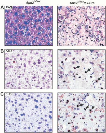
Figure 4.
Apc2Δ/flox Mx-Cre hepatocytes cannot maintain liver function and arrest in mitosis. (A) PAS staining. Most Apc2Δ/flox Mx-Cre hepatocytes were PAS-negative as indicated by the loss of the staining. (B) Ki67 and (C) pH3 staining of Apc2Δ/flox and Apc2Δ/flox Mx-Cre livers at day 11 after pI/C injection. The majority of Apc2Δ/flox Mx-Cre hepatocytes were in mitosis. Arrowheads point to Ki67- or pH3-positive cells.

