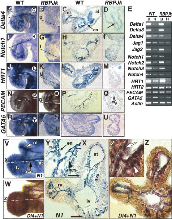
Figure 1.
Endocardial expression of Notch and marker genes. Whole-mount and transverse sections of E8.5–9.5 hearts. (A–D) Delta4 mRNA is expressed in the endocardium of wild-type embryos (A, C), and is severely reduced in RBPJk mutants (B, D). (E) Semiquantitative RT-PCR analysis in E8.5–9.0 wild-type and RBPJk mutants. (B) Body; (H) heart. (F–I) Notch1 is strongly expressed in the endocardium of wild-type embryos (F, H) and is strongly reduced in RBPJk mutants (G, I). (J–M) Endocardial HRT1 expression in wild-type (J, L) is strongly reduced in RBPJk mutants (K, M). (N–Q) PECAM1protein is present in the endocardium of wild-type (N, P) and RBPJk (O, Q) embryos. (R–U) GATA5 is expressed in the endocardium and myocardium of wild-type (R, T) and RBPJk (S, U) embryos. (V–Z) E9.5 wild-type hearts. (V, X, Y) Notch1 expression. (V, Z, Z′) Double in situ with Delta4 (red) and Notch1 (blue). (X, Y) Notch1 expression in endocardium (arrow in Y) and mesenchyme (arrowhead in Y); (Z, Z′) Delta4 and Notch1 coexpression. Coincident signal produces black/purple coloration. Dashed lines indicate plane of section. (en) Endocardium; (ot) outflow tract; (avc) atrio-ventricular canal; (at) atrium; (lv) left ventricle, (rv) right ventricle; (mes) mesenchyme. Note the defective looping of RBPJk mutants. Scale bar, 25 μm in M, O; 100 μm in L, N.

