Figure 4.
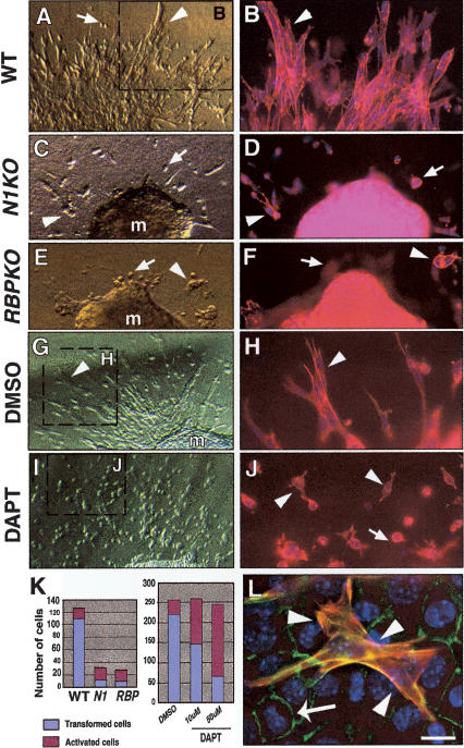
Impaired EMT in Notch1, RBPJk, and DAPT-treated cardiac explants cultures. E9.5 atrio-ventricular canal explants cultured for 72 or 96 h. (A, B) Wild type. (C, D) Notch1 mutant. (E, F) RBPJk mutant. (G, H) Wild-type explant cultured with DMSO. (I, J) Wild-type explant cultured with 10 μM DAPT. (B, D, F, H, J) Phalloidin-TRITC staining. Transformed mesenchymal cells invadingthe collagen gel are indicated by arrowheads, and activated but not transformed cells by arrows. (K) Quantitative analysis of explants. (Left table) The number of transformed mesenchymal cells is significantly reduced in explants from Notch1 and RBPJk mutants. (Right table) Wild-type DAPT-treated explants show a dose-dependent reduction in transformed mesenchymal cells and an increase in activated but not transformed cells. (m) Myocardium. (L) Wild-type explant with transformed cells expressing α-SMA-Cy3 (arrowheads) surrounded by nontransformed endothelial cells stained with Phalloidin-FITC. Scale bar, 15 μm in A, C, E, G, I; 5 μm in B, D, F, H, J; and 2 μm in L.

