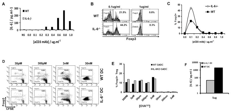
FIGURE 6. IL-6 partially antagonizes low-dose Treg expansion but strong TCR stimulation blocks Foxp3 expression independently of IL-6.
A–C, CFSE-labeled CD4+ T cells from IL-6−/− or WT C57BL/6 mice were stimulated for 5 days with plate-bound anti-CD3 at the indicated doses plus soluble anti-CD28 at 1ug/ml, then stained for CD4 and Foxp3. A, IL-6 production by purified CD4+ T cells from WT or IL-6−/− mice after 48 hours of culture, detected by Luminex. B, Foxp3 expression in gated WT or IL-6−/− CD4+ cells after 5 days stimulation with indicated doses of anti-CD3 plus anti-CD28. C, Induction of Foxp3+ Treg from WT or IL-6−/− CD4+ T cells after 5 days culture with the indicated concentrations of anti-CD3 mAb.
D–F, CFSE-labeled CD4+ T cells from OT-II mice, stimulated for 7 days with G4DC from WT or IL-6−/− mice and the indicated concentrations of OVA323 peptide. D, CFSE and Foxp3 expression in gated CD4+ OT-II T cells after 7 days stimulation. E, Percentage of Foxp3+ OT-II Treg at 7 days. F, IL-6 production, detected in supernatants from co-cultures of OT-II and WT or IL-6−/− DC. All data shown are representative of two independent experiments.

