Figure 1.
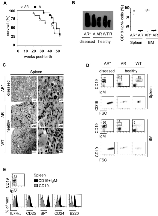
Phenotype of pro-B-cell leukemia in AR mice. AR and A mice were kept in a pathogen-free animal facility and were euthanized at the first signs of illness. (A) Kaplan-Meier analysis of overall survival as a percentage of AR mice (◊, n = 19) and A mice (■, n = 17) without any treatment over a 52-week follow-up period postbirth. A log-rank test was used to compare survival in the 2 cohorts (P < .3887). (B) Macroscopic splenomegaly in diseased AR* mice, compared with spleens of healthy, age-matched A, AR, WT, and R mice, as well as a CD19+IgM− cell population as the percentage of total MNCs in the spleen and BM of diseased AR mice (AR*) and age-matched healthy AR mice. (C) Hematein-eosin staining of spleen sections from an AR* mouse, an age-matched healthy AR mouse, and a WT mouse at 100× and 400× magnification. Lymphoblast infiltration (*) and atypical mitoses (**) are present in the AR* spleen sections. Normal, extramedullar hematopoiesis showing erythroblasts and megakaryocytes is present in the healthy AR and WT mice. (D) FACS analysis of BM and spleen from a diseased AR* mouse and healthy, age-matched AR and WT mice gated on the white blood cell (WBC) populations after RBC lysis. Dot plots indicate the percentage of the immature lymphoblastoid CD19+IgM− (AR*), healthy immature CD19+IgM− (AR), and mature CD19+IgM+ (WT) B-cell compartments in the spleen and the immature (CD19+IgM−) populations in the BM of the indicated mice, respectively. (E) Histograms indicate the surface expression (according to an immunofluorescence analysis) of IL7Rα, CD25, BP1, CD24, and B220 gated on the CD19+IgM− population of an AR pro-B-cell leukemia sample from the spleen.

