
Figure 7.
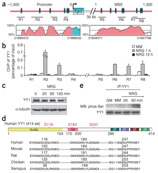
YY1 binds to chromatin at the Egr2 locus only in Schwann cells treated with NRG1. (a) Schematic diagram of the promoter and MSE of Egr2. YY1 consensus binding sequences (green ovals) and the regions (R1–R8) analyzed by chromatin immunoprecipitation are indicated. The plot in pink shows highly conserved sequences between rat and human. Scale marks on x axis indicate 50 bp. (b) Chromatin from Schwann cells cultured in minimal medium (MM, white bar) or treated with NRG1 for 1 h (gray bars) or 12 h (black bars) was immunoprecipitated (ChIP) with antibodies against YY1 and the regions indicated in a were amplified. The results are expressed as percent of input. YY1 was recruited to multiple regions of the Egr2 promoter and MSE after 1 h NRG1 treatment. (c) Protein blot analysis of YY1 in Schwann cells after NRG1 treatment. (d) Schematic diagram of human YY1 protein including the acidic N terminus, the glycine-lysine–rich central domain (GK) and the C-terminal DNA-binding domain, which is composed of four zinc fingers (ZF). Three highly conserved serine residues are marked in red. (e) Co-immunoprecipitation of protein lysates from rat Schwann cells kept in growth medium (GM) or minimal medium (MM), or treated with NRG1 for 20 min or 1 h. After immunoprecipitation (IP) with anti-YY1 antibodies, the protein blot (WB) was probed with anti-phospho-serine antibodies. Full-length blots are presented in Supplementary Figure 5.