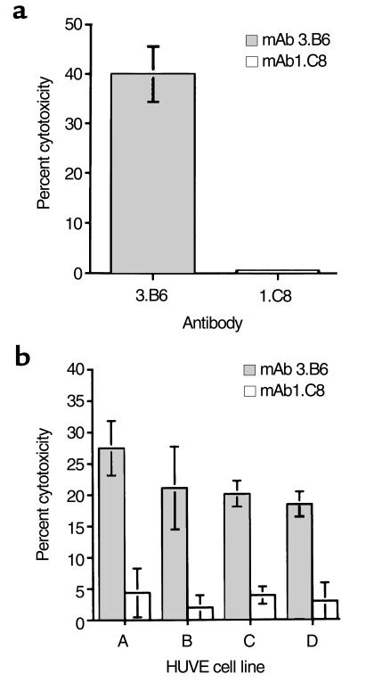
Figure 8.
(a) Cytotoxic activity of human mAb 3.B6 on HUVE cells. HUVE cells were isolated from three umbilical cords and pooled as described (28). Then, mAb 3.B6 and control mAb 1.C8 (human IgM anti-streptococcal/anti-myosin antibody) were tested at 10 μg/mL. Percentage of cytotoxicity was determined by the formula: % lysis = [(test release – spontaneous release)/ (maximum release – spontaneous release)] × 100. Spontaneous release was measured using IMDM without antibody, and maximum release was measured using 1 N HCl. (b) Cytotoxic activity of mAb 3.B6 on four primary HUVE cell lines that were isolated from different umbilical cords. Monoclonal antibody 3.B6 induced cytotoxicity (gray bars) and no cytotoxicity observed by isotype control mAb 1.C8 (open bars). Percentage of cytotoxicity was determined by the formula as in a. Spontaneous release was measured using IMDM without antibody, and maximum release was measured using 1 N HCl.

