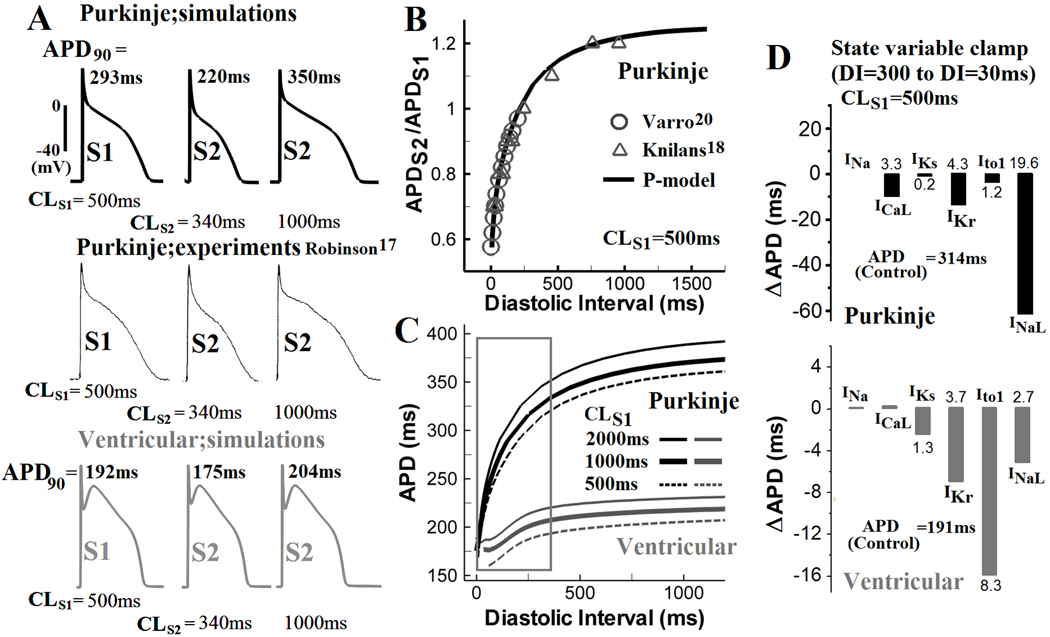
Figure 3.

APD restitution. (A) After steady state was reached during pacing at CL of 500ms (S1), an additional stimulus (S2) was applied at a coupling interval of 340ms or 1000ms from the last S1 stimulus. Simulated Pcell S1 and S2 APs (top) are compared with experimental recordings (middle)17 and with Vcell APs (bottom). (B) Simulated and experimentally measured20,18 Pcell APD restitution curves (CLS1=500ms). (C) Comparison of APD restitution in Pcell (black) and Vcell (gray) for S1 pacing at 500ms, 1000ms and 2000ms. Restitution is steep for S2 coupling interval <300ms in Pcell (box). (D) Top: Changes in Pcell (top) and Vcell (bottom) APD (ΔAPD) when INa, ICaL, IKs, IKr, Ito1, and INaL at diastolic interval (DI) of 300ms were reset to their values at DI of 30ms, for CLS1= 500ms. Bottom: same as top for Vcell.
