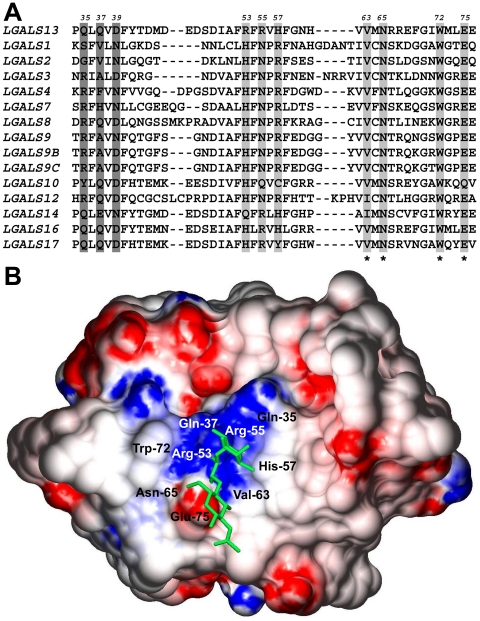
Figure 5. PP13 binds to blood-group H antigen in silico.
(A) Amino acid sequence alignment of 14 human galectins (partial view). Highly conserved residues in the CRDs that are involved in carbohydrate binding are highlighted in light gray, conserved residues in PP13 CRD are denoted with asterisks. B-sites that are involved in blood-group antigen binding are highlighted with dark gray. Amino acid positions in PP13 are shown above the sequences. (B) Surface representation of PP13 complexed with blood-group H trisaccharide (stick representation). Blue and red indicate positive and negative electrostatic potentials mapped to the molecular surface, respectively. As in CGL2, the binding groove of the PP13 CRD contains a central positive channel flanked by negative regions.

