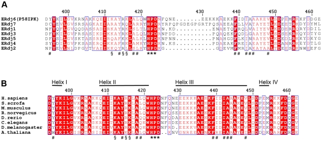
Figure 4. Sequence conservation between J domains from the human ERdj proteins and from P58IPK from different species.
(A) Alignment of sequences of the J domains from the human ERdj proteins. The surface residues mentioned in the text are labelled, using (*) for the residues of the conserved HPD motif, (#) for residues of the adjacent hydrophobic patch in human P58IPK, and (§) for positively charged residues on helix II. Conserved residues are shown with red background, and residue numbering corresponds to human P58IPK. (B) Sequence alignment of the J domains from P58IPK from different species. Labelling is the same as in (A).

